McGill's brain-imaging database a first
McGill's brain-imaging database a first McGill University
User Tools (skip):

McGill's brain-imaging database a first
Technology takes guesswork out of data collection
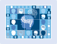
Alan Evans is almost giddy as he counts down to the launch of the world's first online brain-imaging database, which will allow scientists to better understand how the human brain develops.
"I've been at McGill for 23 years, and I don't think I've ever felt this kind of buzz. It's been just a perfect storm of data, people and the Zeitgeist of the field," said Evans, a biomedical engineering professor at the Montreal Neurological Institute and the Principal Investigator of the Data Coordinating Centre for the MRI Study of Normal Brain Development.
To build the database, researchers at six American pediatric study centres collected three-dimensional Magnetic Resonance Imaging (MRI) scans of more than 500 children, from newborns to aged18, who had no neurological disorders. The database will be made available to the worldwide scientific community via a website scheduled to go online in August.
Kids' brains scanned for baseline
TZIGANE
Each child's brain was scanned a minimum of once every two years over a period of six years — more often for younger, more rapidly developing children. The data will provide a point of comparison for researchers interested in observing how a healthy brain develops, as opposed to a brain affected by a neurological disorder such as autism or child-onset schizophrenia.
Such data has never been available before, largely because the technology didn't exist.
"What used to be called fishing is now called data mining. We now have the technology to collect vast amounts of data and incredibly powerful computers capable of handling it," said Evans. "As a colleague of mine said to me, we're not fishing anymore. We're stocking the lake. This changes the whole way of approaching science."
Work on the database started in 2000, after the U.S. National Institutes of Health, funders of the project, decided that existing neurological studies were often too small in scale and tended to rely on "populations of convenience" — the subjects were typically the friends or relatives of researchers.
The MNI was chosen to lead the project because of its expertise in the relatively new field of brain imaging, Evans said.
To ensure the validity of the data, the researchers enlisted Info U.S.A., a marketing agency, to provide a representative sampling of potential candidates using demographic information from the U.S. Census Bureau.
A mailing was first sent out to prospective families asking them if they would be willing to take part in the project. Those families who expressed interest entered the first stage of a rigorous screening process that included interviews and neurological examinations. They would go on to subsequent stages only if they met all the demographic, medical and other selection criteria. Children were excluded if their mothers smoked more than half a pack of cigarettes a day during pregnancy, for instance. In two cases, the children were removed from the project and immediately referred to a pediatric neurologist after they were found to have major structural brain abnormalities.
The recruitment process was anything but easy, Evans admitted.
"We had to convince parents to bring in their children, who had nothing wrong with them, to get their brains scanned. Getting them all to agree was a hell of a job. There was a lot of cold-calling."
The result of all this time and effort is a unique collection of downloadable images and behavioural data mapping the physiological changes that occur as healthy, normal brains develop.
"This will allow work to be done that was inconceivable in the past," said Bradley Peterson, director of MRI research at Columbia University and the New York State Pyschiatric Institute, who was among the advisors who helped set the parameters of the database project. "Understanding how things go right helps us to understand how they go wrong. When you're studying patients with attention deficit hyperactivity disorder, obsessive compulsive disorder, anxiety or depression — all the important conditions in childhood — having a set of control images of normal brain development will be extremely valuable."
The total budget is about $30 million, of which $9 million funded the work done at McGill, where the software program that runs the database was designed. The other institutions involved in the project are the Children's Hospital in Boston, the Cincinnati Children's Hospital Medical Center, the University of Texas Houston Medical School, the Neuropsychiatric Institute and Hospital, the University of California Los Angeles (UCLA), the Children's Hospital of Philadelphia and Washington University in St. Louis, Missouri.
Once the database is officially "released" for use by scientists the world over in August, McGill and the MNI will be credited more and more frequently as sources of expertise for the most important breakthroughs in neurological research. "We could be analyzing this data for the next 50 years," said Evans.